Villard Tube (ca. 1900)
Ref. B6


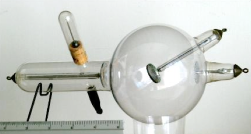
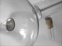
This is a ten-inches (25 cm) long, four inches (10 cm) bulb. Concave aluminum cathode. Platinum anti-cathode (with a visible central focal spot). Flat disc anode. Made by Emil Gundelach, bearing the DRP (Deutsches Reichspatent) No. 103100 dating back to 1898.
Basically similar in design to the “Bulbar Focus Tube” described earlier, with the addition of a regeneration device using Paul Villard’s osmotic method.
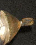
Note the platinum wire electrode terminals looped around the neck of the small copper dome-shaped external connections of the tube (picture above, right).
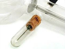
This regeneration device, introduced by Paul Villard (1860-1934) consists of a small capillary platinum (or palladium) tube, closed at the external end while the other end is sealed through the glass wall and communicates with the cavity of the X-ray tube. With use the gas pressure inside the tube drops down and the tube becomes “hard”, needing a higher voltage to operate. By heating the external part of the capillary in a flame, gas diffuses through the capillary into the tube and restores the correct pressure. The external part of the regeneration device is usually seen protected by a small glass test-tube.
Tube donated by the late Rev. François Dupré-Latour, sj, ex-Dean and Professor of Physics at the Medical School of the St. Joseph University in Beirut. It had belonged to the late Rev. Maurice Collangettes, sj, Professor of Physics during the early years of the 20th century, and who had received from France, in 1900, his first X-ray outfit.