Pear Shaped Crookes Tube
Ref. A2
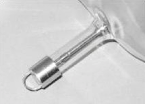
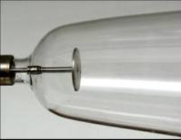
Instead of having the electrodes “in line” on the opposite sides of the tube, as in the early Crookes tube, the cathode here is a flat aluminum plate on the narrow side of the “pear” while the anode is “rod shaped” in a secondary lateral tubular chamber on the larger side. The other tubular extension is for the vacuum.
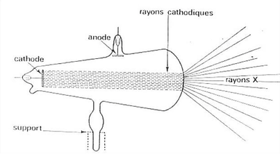
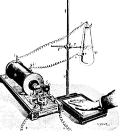
It is while experimenting with a similar tube that Röntgen incidentally discovered X-rays late in 1895. These rays, of an unknown nature yet, were produced by a large area of the glass wall on the wider side of the tube, by the impact of the unfocused “cathode rays” (electron beam). This large size of the X-ray source accounts for the lack of sharpness of the earliest X-ray “photographs”.
Such tubes were soon abandoned for the production of X-rays, and replaced by what came to be known as “the focus tube”.
This particular tube is a late 20th century replica, offered by Siemens.