Philips 9” Image Intensifiers
Ref. J9


The first image intensifiers in Radiology were introduced by Westinghouse (the Fluorex ) in 1952, followed by the Philips intensifier, and by the French made “Fluoricon” of General Electric by “Thomson-Houston, France”. These early image intensifiers had a 5” (12.5 cm) input screen, and their intensification factor was low, well below 1000. Then came the 7” (17.5 cm) intensifier by Siemens, and the 6” (15 cm) by Philips, with improved characteristics. A race for larger intensifiers followed, and in the early Seventies, Siemens introduced the largest intensifier ever made, with a 23” (57 cm) input screen but it was soon abandoned, reportedly because of some accidental implosions.
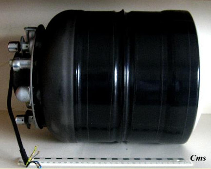
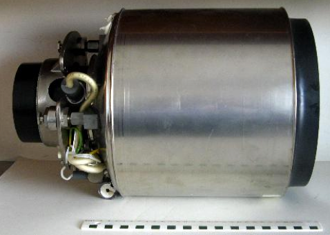
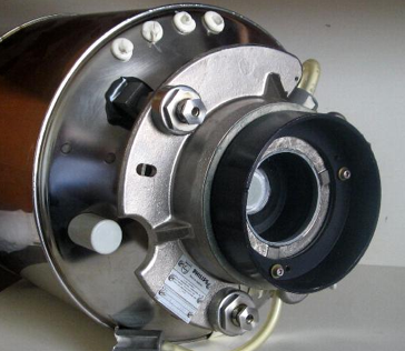
The above-pictured tubes dating respectively to the mid-eighties and late nineties of last Century show some physical differences but have highly similar technical specifications, with an amplification factor of over 1000. The anterior input fluoroscopy phosphor measures about nine inches in diameter while the output phosphor in the back is only about one inch. The bright image on the output phosphor was transmitted to a TV monitor by a TV camera tube through an elaborate optical system that has later been replaced by fiber-optics. Latest technology transmits the intensified image from the output phosphor through fiber-optics to a CCD sensor (charged coupled device) or to other type sensor, as found in digital photo cameras.
Picture adapted from “Introduction to Medical Radiographic Imaging”, Eastman Kodak Company, 1993, p162.