Speakers and Abstracts
(Speakers are listed in the order of the programs’ sessions)
Lecture 1: TAVI: Kaleidoscope of Cases & Devices. Interactive Panel Discussion
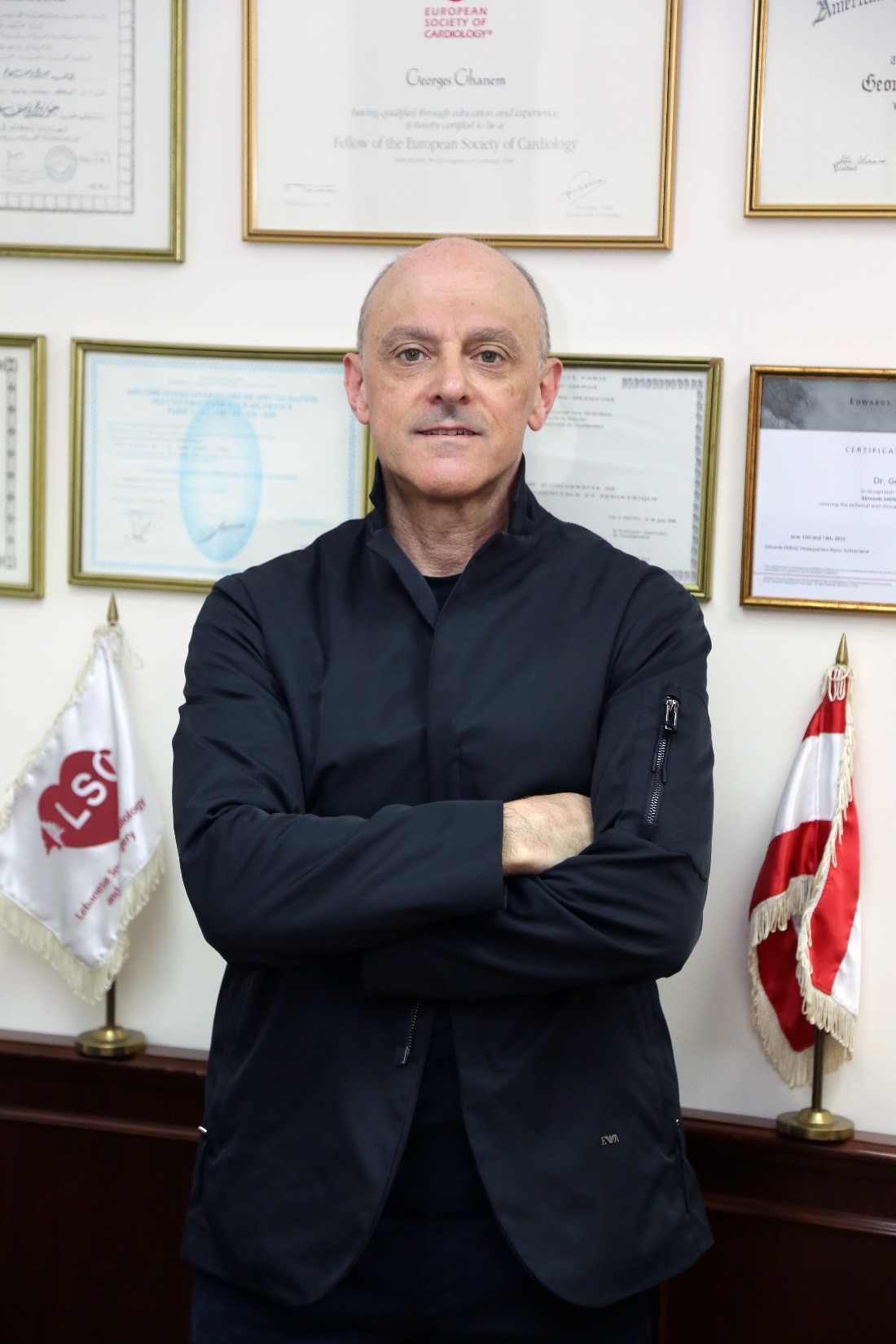
Georges Ghanem, MD, FESC, FACC
Dr. Ghanem obtained his MD degree from Saint Joseph University in Lebanon in 1985. He then pursued his Cardiology Residency & Fellowship in Paris followed by multiple training in USA.
He has been appointed as Fellow of the European Society of Cardiology in 2006 and Fellow of the American College of Cardiology in 2007. He was also elected as President of the Lebanese Society of Cardiology (LSC) in 2005.
In his clinical practice, Dr. Georges Ghanem was a pioneer in introducing major interventional cardiology techniques in Lebanon since 1993.
His name appears in more than 70 publications.
In 2019 he achieved a professional certificate on strategic health planning and in 2020, a professional certificate on Management and Leadership at the International Center of Parliamentary Studies in London.
In September 2016, he was appointed as Chief Medical Officer at the LAU Medical Center-Rizk Hospital.
In September 2017, he was promoted to Full Professor in Cardiology at LAU Gilbert and Rose-Marie Chagoury School of Medicine.
In February 2023, he was appointed as Deputy CEO for Strategy & Development at LAU Medical Center-Rizk Hospital.
In parallel to his activities in the medical field, Dr. Ghanem is a Master in Martial arts; he pursues an intense activity in many sport techniques and conduct counselling activities in sport & health domain. He has also an extensive experience in war medicine.
He is a founding member of “The White Coat Organization” addressing the issue of corruption and the incompetence of the authorities to control the healthcare crisis. He launched a movement: The National Assembly for Lebanon Identity.
During the Covid pandemic, Dr. Ghanem played a national role with the LAU mobile clinic, raising awareness, performing massive PCR testing and an aggressive vaccination campaign on all the Lebanese territory.
Abstract
During this talk, interactive clinical cases addressing multiple challenges in TAVI patients will be presented. This will highlight the importance of doing almost all patients with femoral approach where data revealed striking evidence of superiority compared to all other approaches.
The state of the art of doing ring fracture for valve in valve will be shown.
The importance of preserving coronary access in challenging coronary origin will be tackled.
Other challenging clinical settings (kyphoscoliosis, large bicuspid highly calcified annulus, valve with previous metallic mitral valve replacement…) will be discussed.
The feasibility of TMVI will be also presented and discussed at the end.
The audience’ opinion will be also assessed through few questions with direct voting.
Lecture 2: Key Elements to Decide Between BEV vs SEV: Is there any Evidence?
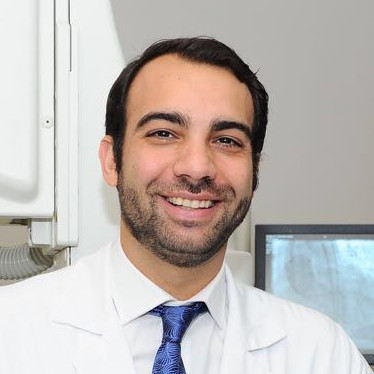
Fadi Sawaya, MD, FACC
Dr. Sawaya is an Assistant Professor of Internal Medicine in the Division of Cardiology at the American University of Beirut Medical center (AUBMC). Dr. Sawaya received his Medical Degree with Distinction from the American University of Beirut in 2008 and was the recipient of the Alpha Omega Alpha awards. After which, Dr. Sawaya completed 6 years of residency and fellowship training in Internal Medicine and General Cardiology at Emory University hospital in Atlanta (2008-2014). He then served as the Chief Interventional Cardiology Fellow for one year at the Andreas Grüentzig Cardiovascular Center at Emory University (2014-2015). After his time in the United States, Dr. Sawaya moved to Europe to the Institut Cardiovasculaire Paris Sud, France and Rigshospitalet, University of Copenhagen, Denmark, two of the most experienced and highest volume centers in Europe, where he did a clinical Fellowship in Complex Coronary Interventions, Endovascular, Structural and Adult Congenital Heart Interventions for 1.5 years.
He is currently the Director of the Structural Heart program at AUBMC and his clinical interest is performing TAVI, mitral Clip, LAA occlusion, Paravalvular leak closure and ASD/PFO closures and CTO interventions.
Abstract
One crucial decision in TAVR procedures is the selection between self-expandable and balloon-expandable transcatheter heart valves (THVs).
The choice between self-expandable and balloon-expandable THVs involves a meticulous evaluation of patient characteristics, anatomical considerations, procedural intricacies, and device-specific attributes.
This presentation synthesizes current evidence, clinical guidelines, and expert consensus to provide a comprehensive framework for decision-making in the selection of transcatheter heart valves. By understanding the differences of self-expandable and balloon-expandable THVs and their appropriateness in various clinical scenarios, clinicians can optimize patient outcomes and enhance the delivery of transcatheter interventions in structural heart disease.
Lecture 3: TAVI in Moderate Aortic Stenosis: A Game Changer

Georges Ghanem, MD, FESC, FACC
Abstract
- Moderate AS may not be as benign as we thought and is associated with increased mortality.
- AS may not only affect the valve but also a complex myocardial response & damages.
- Markers of this damage could be the LVF, LVH, LA dilatation, GLS, Fibrosis …
- Symptoms are the clinical marker of these damages.
- Some of these damages are irreversible once AS become severe & symptomatic
- Some unknowns: Lpa & LPA genomics which may identify a high risk sub-group in moderate AS patients.
- Accurate measurement parameters (lvot, low flow, low gradient…); DSE; CAC Score; Hemodynamic & cath… important tools to perform an accurate & reproducible diagnosis.
- In 1968, the landmark publication by Braunwald & Ross in Circulation, highlighted the steep decline in survival in patients with severe AS at the onset of symptoms.
- EXPAND II - TAVR UNLOAD - PROGRESS Could they change this evidence?
- A role for artificial intelligence in predicting faster disease progression in moderate AS patients?
- Instead of waiting for symptoms to arise before intervening, should we pivot towards detecting myocardial damage to inform our management strategy in Moderate AS?
Lecture 4: MitraClip 5 years Follow-up: Confirming Coapt or shifting to Mitra FR Trend?
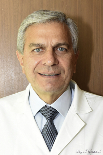
Ziyad Ghazzal, MD
Dr. Ghazzal earned his MD degree from AUB Faculty of Medicine and completed his residency in internal medicine at AUBMC in 1984. Dr. Ghazzal has upheld an academic rigor in all his professional life, becoming an internationally renowned interventional cardiologist who has excelled as a highly accomplished clinician, researcher, and teacher/ mentor. He was promoted to Professor of Medicine at Emory University, Atlanta, GA, where he has been a full-time faculty member since 1990, after completing his fellowship training at the same institution from 1985 to 1990.
Dr. Ghazzal has published extensively in the cardiology literature, including high-quality original papers and many other reviews, editorials, book chapters, and video seminars. He is regularly sought after by international organizations for clinical lectures, tutorials, and other CME activities in the area of interventional cardiology. As a director of the Interventional Fellowship Program at Emory, Dr. Ghazzal has played an instrumental role in the success of the training program there, one of the premier programs in the United States. He has been a member of a large number of prospective multicenter clinical trials, including being the local principal investigator at Emory. He was an associate editor of the popular ‘Cardiosource’, the official teaching website of the American College of Cardiology. He was also the co-chair of the international distant learning program of the College. He is an editorial consultant for JACC Cardiovascular Interventions and the American Journal of Cardiology.
Dr. Ghazzal is an Alpha Omega Alpha member and has received many honors, including the Medical Leadership Award from the National Association of North America, and the Excellence in Teaching Award from Emory University. In 2008 Dr. Ghazzal joined the American University of Beirut where he functioned as the Deputy to the Executive Vice President/Dean of the Faculty of Medicine and Medical Center in addition to his duties as an interventional cardiologist. He was appointed interim Medical Center Director and Chief Medical Officer (2017-2020). He is the Founding Director of the Heart & Vascular Institute and currently serves as the Director of the Moufid Farra Heart, Vascular & Kidney outpatient center.
Abstract
The classic indication of transcatheter mitral valve repair with the MitraClip has been for the reduction of significant symptomatic mitral regurgitation (MR grade ≥3+) due to a primary pathology in the mitral apparatus referred to as degenerative MR, when the risk of mitral valve surgery has been determined as prohibitive.
In 2019, the U.S. Food and Drug Administration approved the MitraClip for patients with normal mitral valves who develop heart failure symptoms and moderate-to-severe or severe functional mitral regurgitation due to a reduced left ventricular ejection fraction, despite optimal medical therapy. This approval relied primarily on the randomized COAPT Trial (Cardiovascular Outcomes Assessment of the MitraClip Percutaneous Therapy for Heart Failure Patients With Functional Mitral Regurgitation), which enrolled 614 patients and demonstrated a significant response of functional regurgitation with MitraClip. The clip resulted in a lower rate of hospitalization for heart failure, lower mortality, and better quality of life and functional capacity within 24 months of follow-up than medical therapy alone. MR severity remained moderate or lower in 95% of patients at 1 year. The prespecified goal for freedom from device-related complications was met. At five years, transcatheter edge-to-edge repair of the mitral valve was safe and led to a lower rate of hospitalization for heart failure and lower all-cause mortality than medical therapy alone.
In the MITRA-FR trial, more than 300 HF patients with severe MR were randomized to medical treatment alone or with MitraClip. At one year, the risk of death or the risk of hospitalization for heart failure were similar in both groups.
It is important to select the appropriate candidates for MitraClip in heart failure patients with secondary mitral regurgitation. Currently, ideal patients are those presenting with chronic CHF and severe secondary MR with a reduced EF (20-50% but preferably 20-35%). Patients should be on optimal medical therapy for heart failure and categorized as high surgical risk with mitral valve surgery but with anatomically suitable mitral valve anatomy for MitraClip. Patients with ischemic cardiomyopathy should not have significant residual ischemia from potentially revascularizable territories. It is crucial to properly assess the mitral regurgitation by resorting to accepted gold standards and evaluate the possible benefit from cardiac resynchronization therapy. It is also important to factor into the decision other important considerations such as the presence of irreversible pulmonary hypertension or severe right ventricular dysfunction. Treatment decisions should be individualized through a heart team approach while combining clinical findings with multi-image modalities as required.
Lecture 5: MITRA-FR vs. COAPT What we Know Now: A case Presentation
Richard Zalloum, MD
Dr. Zalloum is a Clinical Assistant Professor in the Department of Internal Medicine, Division of Cardiology at the LAU Gilbert and Rose-Marie Chagoury School of Medicine and LAU Medical Center-Rizk Hospital.
Abstract
Managing functional mitral regurgitation (MR) poses significant challenges, primarily due to its strong association with left ventricular (LV) dysfunction. Therapeutic approaches predominantly focus on addressing the underlying LV disorder. Strategies involving neurohormonal antagonists and resynchronization therapies have shown promise in improving outcomes by reducing left ventricular volumes. Conversely, interventions aimed at the mitral valve have produced conflicting results, underscoring the importance of reversing LV dysfunction in this patient cohort. Recent evidence from the COAPT and MITRA-FR trials highlights the heterogeneous nature of functional MR patients which could account for such conflicting observed data, categorizing them into those with MR predominantly caused by global mitral valve distortion due to LV enlargement and those with LV pathology disproportionately affecting segments supporting normal mitral valve function. These aforementioned trials suggest that patients with disproportionate MR may benefit more from mitral valve-targeted interventions. The question arises: which functional MR patients can benefit from mitral valve interventions, and how can we identify them? We present a case of a 55-year-old female admitted to our emergency department in 2019 with myocarditis, complicated by depressed systolic function (EF 30-35%) and severe mitral regurgitation. Despite refractoriness to multiple vasopressors, the patient underwent a MitraClip intervention following shared decision-making, guided by evidence from the COAPT trial.
Lecture 6: TS LHC Technique From Basic to Structural Heart Diseases Interventions

Georges Ghanem, MD, FESC, FACC
Abstract
It’s an in deep review of the basics of trans-septal left heart catheterization described by John Ross in 1959 but was in decline after the direct coronary and left heart catheterization were developed by Sones and Judkins. It regain a preponderant position with the mitral balloon valvuloplasty in the 80s and what followed in the advancement of structural heart disease interventions and ablation.
The outline of the lecture will be as follows:
- Historical review
- Embryology & Anatomy
- Technique & Guidance: Fluoroscopy – TEE - ICE
- Complication
- PMC
- Mitra Clip
- LAA closure
- TMVI
Lecture 7: Invasive Cardiac Electrophysiology Interventions in the Hybrid OR
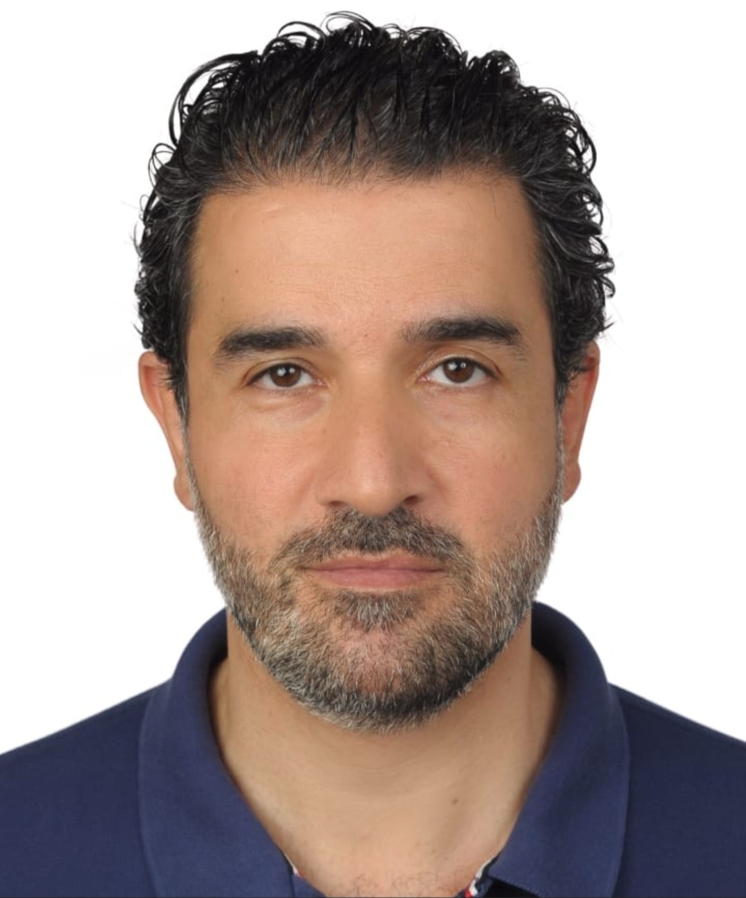
Johny Abboud, MD
Dr. Abboud is Clinical Assistant Professor in the Department of Internal Medicine, Division of Cardiology at the LAU Gilbert and Rose-Marie Chagoury School of Medicine and director of the Cardiology Fellowship program since 2021.
He has a diploma of cardiac electrophysiology and a diploma of cardiac ultrasound from the University of Paris.
Dr. Abboud has more than 15 years experience in invasive cardiac electrophysiology interventions from the implantation of electronic intra-cardiac devices to ablation of complex cardiac arrhythmias.
Abstract
In this presentation, the different electrophysiology procedures conducted in the hybrid OR from the implantation of different types of pacemakers and defibrillators to the extraction of these devices with the potential complications will be discussed.
Also, the different ablation procedures using conventional ablation, 3D mapping and pulsed field ablation highlighting the benefits of the hybrid OR during these procedures will be highlighted.
Lecture 8: Modern PCI in Complex Anatomy/CHIP
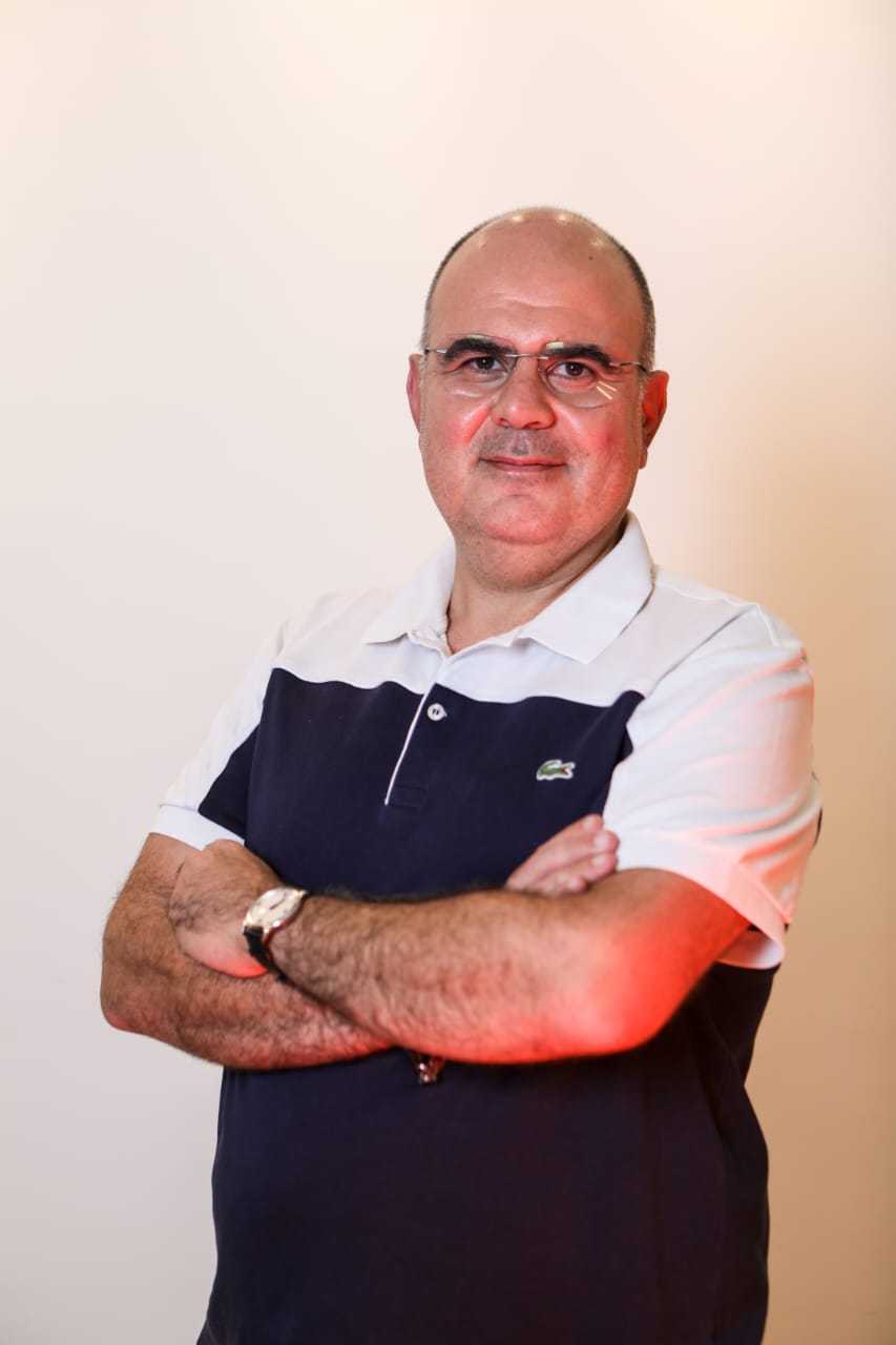
Charbel Naim, MD
Previous Fellow of Interventional & Structural Cardiology at Milan’s San Raffaele Hospital/Antonio Colombo and Centre cardiovasculaire du Centre Hospitalier de l’Université de Montréal.
Dr. Naim accomplished, authored and co-authored original research published in peer-reviewed journals (JACC, CCI, EuroIntervention, JACC-CI, AJC, AJP-Heart and circulatory physiology, CJC,…) and presented at major conferences with several awards.
Currently Assistant Professor of Clinical Medicine at the LAU Gilbert and Rose-Marie Chagoury School of Medicine and practicing Interventional & Structural Cardiologist.
Dr. Naim has particular interest in intravascular imaging and management of complex coronary pathologies in higher-risk patients.
Abstract
The success story of PCI since 45 years wouldn’t be so unprecedented if we did not strive for continuous improvement and innovation.
Our practice in treating higher-risk indicated patients incorporates new technological breakthroughs encompassing modern procedural and intravascular imaging tools, intra-procedural physiologic testing, and the latest lesion preparation and plaque modification tools.
Outfitted with an armamentarium of new techniques and devices we work in consortium with a highly trained team of technicians, nurses and interventionalists with both exceptional technical and cognitive skills to safely achieve adequate revascularization and tailor each patient’s needs.
Lecture 9: Hybrid Approach for Ilio-Femoral Occlusive Disease
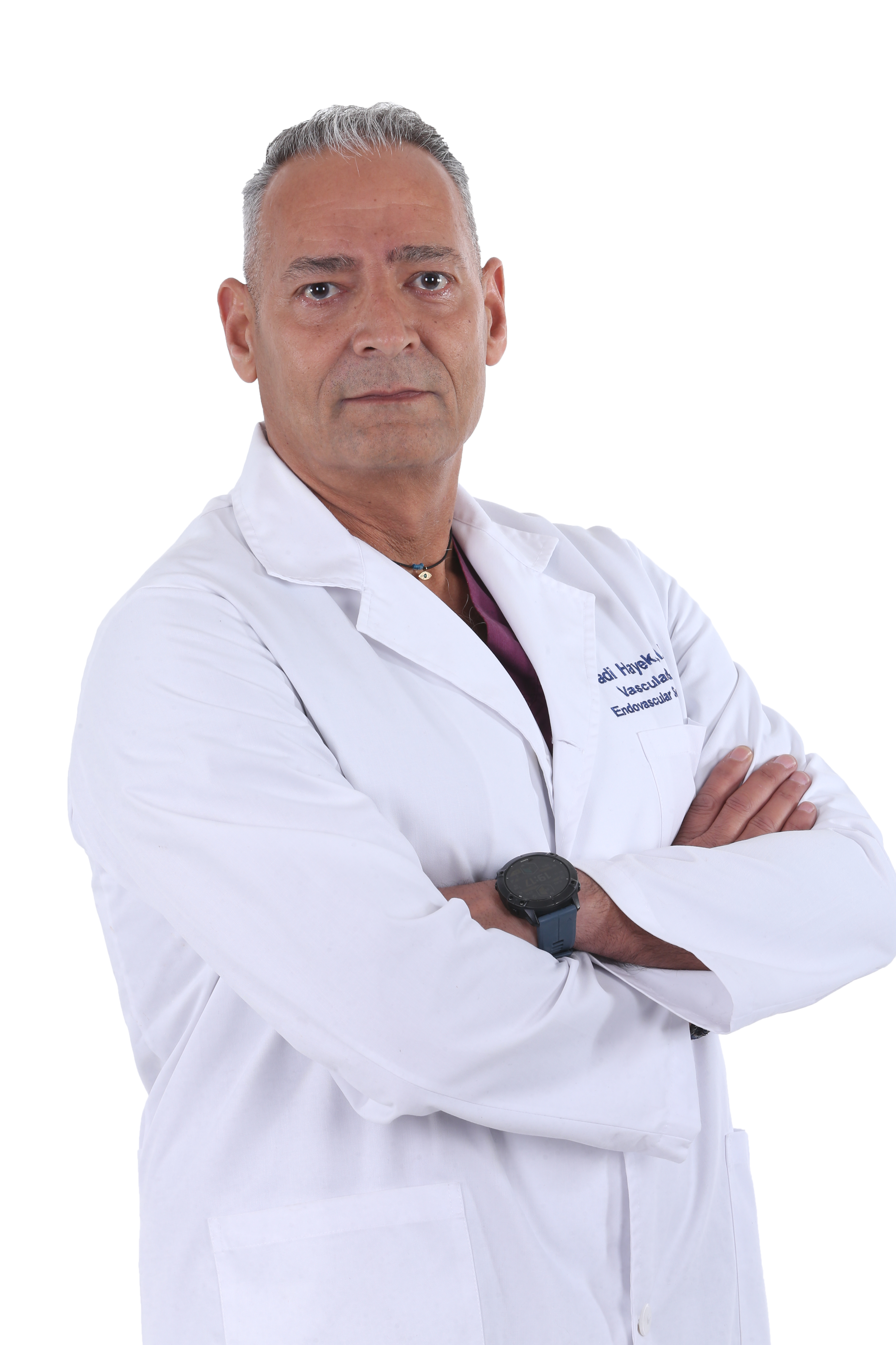
Fadi Hayek, MD
Dr. Hayek is Clinical Associate Professor in the Division of Vascular Surgery at the LAU Gilbert and Rose-Marie Chagoury School of Medicine. He is also the Head of the Division of Vascular Surgery at the LAU Medical Center-Rizk Hospital.
He is the former President of the Lebanese Society for Vascular Surgery (LSVS).
Dr. Hayek received his MD degree from the Saint Joseph University in Beirut in 1988. He then went to France (Bordeaux) where he completed his general and vascular surgery residency and fellowship at the Universite Bordeaux Il in 1996. Dr. Hayek was then appointed as ancien interne et chef de clinique au CHU Pellegrin in Bordeaux, France. Dr. Hayek started his practice in Lebanon in 1996 so accumulating around 28 years of experience in vascular and endovascular surgery. He has shown expertise in vascular and endovascular approaches to aortic surgery (AAA, occlusive disease), limb salvage procedures, carotid surgery, vascular trauma, diabetic foot and the treatment of venous disease and deep vein thrombosis. In addition, he has developed a love and fine expertise in treating and dealing with the management of venous disease. Additionally, he is actively involved in teaching and training medical students and residents in Surgery at the LAU School of Medicine.
Moreover, Dr. Hayek is member of numerous prestigious vascular societies, such as the European vascular society, the French vascular society and the Lebanese vascular society. He presented and has been an invited lecturer at both national and international venues. And, has various surgical publications.
Abstract
Constantly increasing number of procedures performed - endovascular or hybrid in patients with aortoiliac occlusive disease during the last decades finds its explanation in the lower morbidity and mortality rates, compared to bypass surgery.
Hybrid interventions have become an integrated part of our strategy for limb salvage in patients with multi-level arterial occlusive disease.
In this presentation, the commonly used hybrid interventions will be described and their indications and outcomes will be reviewed. TASC II type D aorto-iliac occlusive disease with common femoral artery involvement by hybrid methods of revascularization -iliac stenting with femoral endarterectomy, in patients with multiple medical comorbidities will be reported and the initial experiences of a single center and issues that arose will be discussed.
Lecture 10: Management of Acute Type B Aortic Dissection
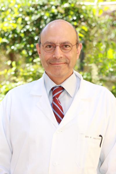
Fady Haddad, MD, FACS
Dr. Haddad is Professor of Surgery at the American University of Beirut (AUB) and specializes in Vascular and Endovascular Surgery. Currently, he is the Head of the Vascular Surgery Division, coordinator of the Endovascular Program and Director of the Non-Invasive Vascular Laboratory. In Addition, Dr. Haddad is the General Surgery Residency Program Director, and was instrumental in the ACGME-I reaccreditation of the program.
He received his training in Vascular Surgery and Transplant Surgery at Boston University and the Boston VA Medical Centers and joined the Cleveland Clinic in 2004-2005, as for his fellowship he completed it in Endovascular Surgery under the mentorship of late Roy Greenberg.
Dr. Haddad has multiple publications in peer review journals on Endovascular Surgery with special interest on EVAR’s and T-EVARs, Carotid surgery and Dialysis access. His practice interest is in endovascular aneurysm repair (Thoracic and Abdominal), Diabetic foot and endovascular revascularization, Dialysis access and modern varicose vein interventions. His current research focuses on prevalence of AAA in the ME region and Aortic Coarctations. He is also the recipient of the 2022 Gold Humanism Honor Society (GHHS) award.
Abstract
Index case: 58-year-old, male patient, known for hypertensive, chronic kidney disease, stable creatinine 2.9 on presentation, presented with acute stabbing interscapular chest pain radiating also to the lower abdomen. He was admitted originally to the CCU and managed with strict blood pressure control, a CT-scan, back then confirmed the presence of type B-aortic dissection starting right at subclavian artery. No significant aortic aneurysmal dilatation, no organ ischemia originally, with normal motor and sensory functions to lower extremities. The patient did well and after 10 days was discharged with normal blood pressure.
He came back with another hypertension crisis about 3 weeks post discharged with again a severe chest and back pain. Again, CT-scan was repeated and showed some extension of the dissection down the Right common iliac with area of very sluggish flow. In addition, more expansion of the false lumen with compression of the true lumen. Slower nephrogram on the Right. At this point, the patient was kept at the hospital and a decision was made to proceed with intervention in his Dissection. The procedure was started with the extra anatomic carotid subclavian bypass and following couple of days later with the stenting of the aortic dissection from the very edge of the left common carotid artery covering the subclavian artery till the distal descending thoracic aorta. Techniques of the procedures will be discussed in the presentation.
There was an immediate perfusion and good expansion of the true lumen, at the end of the procedure, so no further distal stenting, and no further dissection stent or “Petit Coat” extension was done.
The patient did extremely well post operatively and was discharged home with his blood pressure medications. The purpose and the aim of this presentation, and the take home messages, will discuss:
Indications and timing in treating type B dissection.
The value of using a hybrid OR and Hybrid procedures in aortic dissection.
The role and the need to have distal extension with specific dissection stents
Techniques, and the value of IVUS
Lecture 11: Using CO2 for EVAR, Renal Artery Stenting, Is It Worthwhile?
Fadi Hayek, MD
Abstract
Carbon dioxide (CO2) is an excellent negative contrast agent which has been used for a variety of vascular interventions since the introduction of digital subtraction angiography. Due to its high solubility rate and rapid diffusibility via the lungs, CO2 is safe for intravascular usage.
Based on some “live in a box cases” = EVAR, VISCERAL ARTERY STENTING, SFA REVASCULARISATION, using the CO2 angiography, in patients with CKD, we will discuss the indication, side effects, tips and tricks with a review of the literature and clinical evidence.
EVAR under CO2 angiography is feasible, effective, and safe in CKD patients.
Successful outcomes with minimal complications.
Suggests CO2 angiography as a viable option for EVAR or Endovascular procedures in CKD patients.
Lecture 12: How to deal with EVAR when it Goes Wrong?
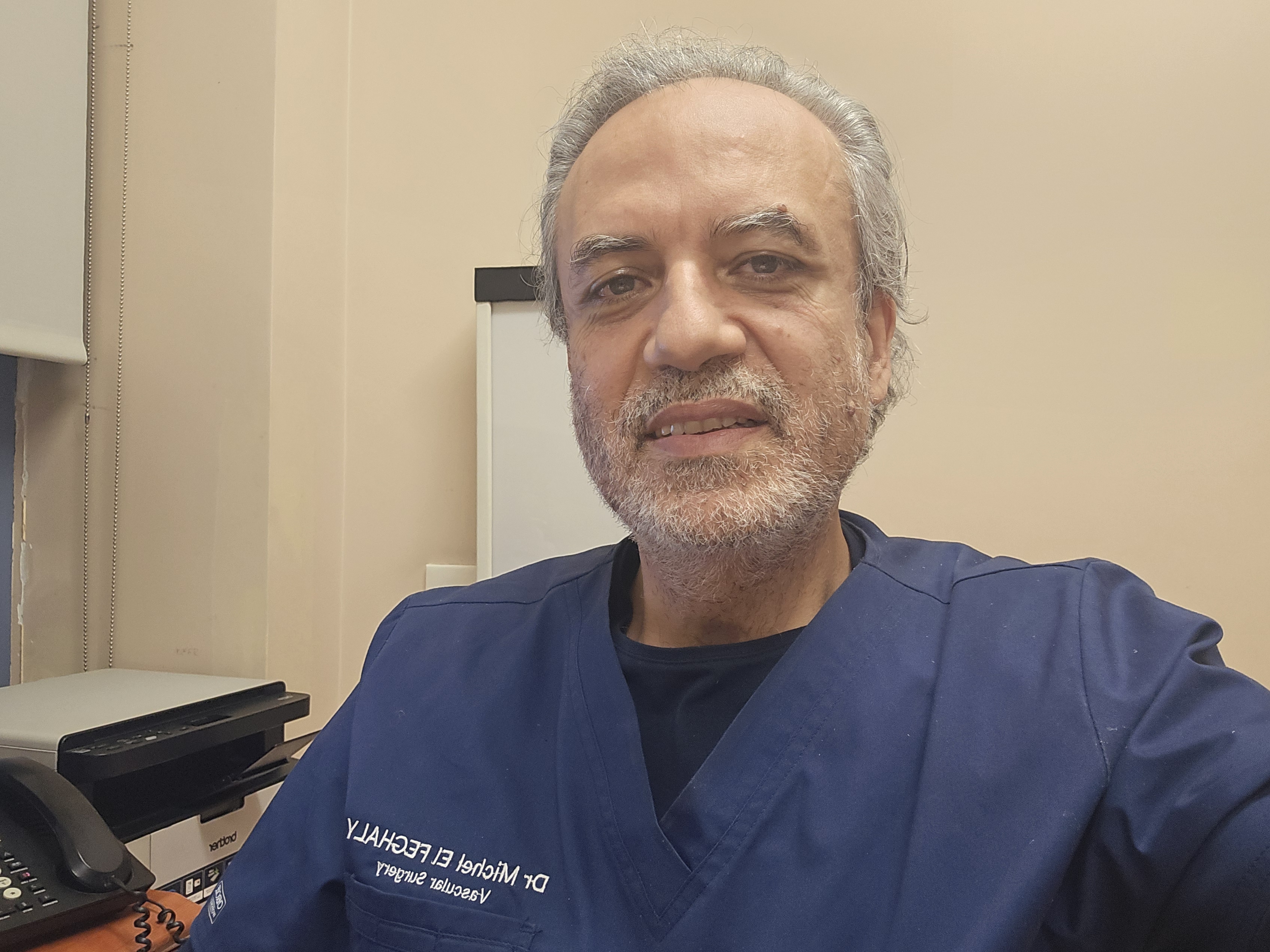
Michel El Feghaly, MD, MBA, FACS
Dr. El Feghaly is Professor in Vascular Surgery. He is currently Head of Division of Vascular Surgery at the Saint George Hospital University Medical Center and Director of the Wound care and training center at SGHUMC.
Abstract
Endovascular repair of infra-renal aortic aneurysm is becoming the first choice method of intervention.
- Contralateral gate cannulation is an important, challenging step during infra-renal aortic aneurysm endovascular repair.
- Maldeployment of the contralateral stent is a difficult situation that mandates urgent intervention.
- We present an interesting case of an endovascular salvage method instead of converting to open surgery.
Lecture 13: Bypass in the Leg after Failed Endorepair: Where do we stand after up to 10 years of follow-up and what was the amputation rate after, is it worthwhile?
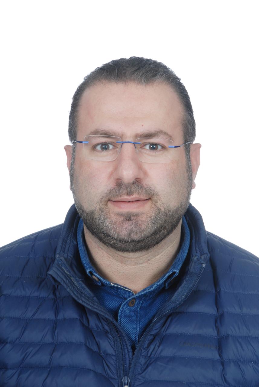
Julien Sfeir, MD
Dr. Sfeir is Clinical Assistant Professor in the Division of Vascular Surgery at LAU Medical Center-Rizk Hospital. He is also the President of The Lebanese Society For Vascular Surgery (LSVS).
Abstract
Objectives: All recent data suggest that percutaneous transluminal angioplasty (PTA) may be appropriate primary therapy for critical limb ischemia (CLI) even in infrapopliteal lesions. In our practice and in selected patients (Lesions type, comorbidities and operative risk) we are treating patients with CLI by endovascular approach. Between 2011 and 2014 and in 24 high risk patients in whom primary PTA was the selected treatment, a conversion to distal Bypass surgery was done due to a failure of PTA and after having complete consent from the patients
Now we evaluate up to 10 years of follow up were do we stand comparing to primary bypass in terms of patency and amputation rate.
Methods: Between 2010 and 2014, 24 high risk patients (Ejection fraction varied between 30 and 40%) selected for primary PTA,were operated for distal venous Bypass for CLI. Conversion to Bypass was due to a failure of recanalization of the lesion and to a complication of the procedure like extensive dissection or arterial rupture.
The median age was 71 years, all patients were diabetic, 2 patients had severe renal impairment under hemodialysis, five patients suffered from COPD. The M/F ratio was 19 men versus 5 women. Operations were done under general or regional anesthesia. All patients were on the same medical treatment (Statin, dual antiplatelet and aspirin)
Results: 24 distal Bypass were done divided as follows :11 in situ Bypass, and 13 by inverted GSV Bypass (11 to Proximal posterior tibialis,2 to pedal artery, 3 to distal posterior tibialis, 3 to Anterior Tibialis, and 5 to Tibio-Peroneal Trunk). The mean operating time was 220 min.Follow up time average was at 18 months. Primary patency at 1 year and at 18 months were respectively 75% and 67%. Secondary patency also at 1 year and 18 months were respectively 80% and 75%. Limb salvage at one year was approximately 80 %. Mortality at 30 days was 12.5% (2 patients died of myocardial infarction and 1 died of respiratory failure due to severe pneumonia). At 18 months the survival rate was around 70%.
Over the 21 patients , 4 died after 3 years and 1 was lost from follow up.
16 patients followed up to 10 years and the results were as it follows:
- 6 occluded their bypasses with 5 minor amputations and 1 occluded with major amputation.
- 9 remained patent with secondary angioplasty or endarterectomy with patch (5 iliac angioplasties with stenting, 1 bypass angioplasty, 3 endarterectomies on the iliofemoral junction).
- Patency rate was 56 % at 10 years.
- Minor amputations was 31 % mostly in the occluded group.
- 1 major amputation (patient had COVID and arterial thrombosis who couldn’t be operated).
Conclusions: When primary amputation is the only remaining option and after failed attempts of endovascular revascularization, and even in high risk patients, surgical bypass must remain an option with a good average of survival, patency rates, and limb salvage even at 10 years of follow up with an optimal medical treatment.
Lecture 14: Ruptured AAA in a high-risk patient with Complex Anatomy for EVAR
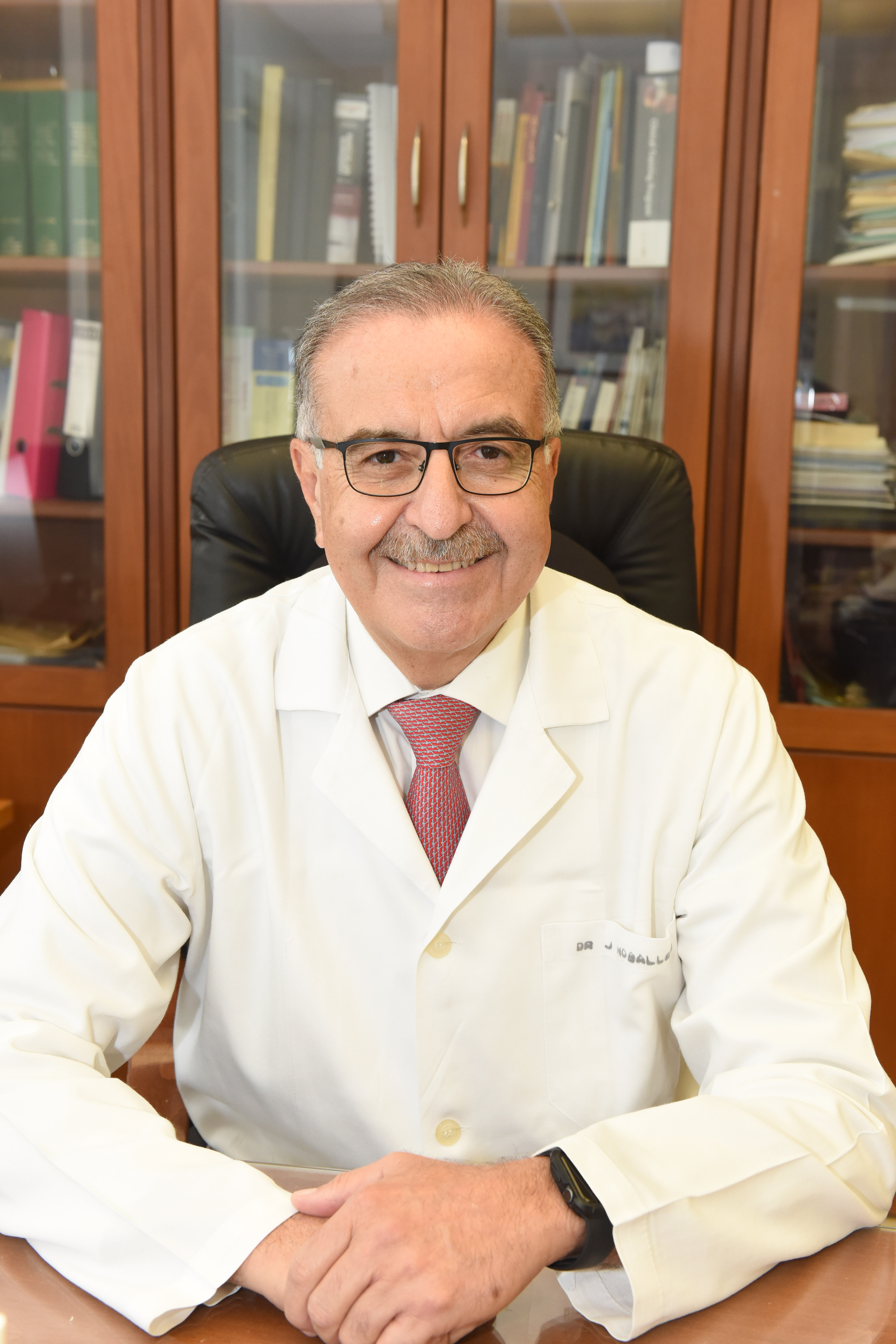
Jamal Hoballah, MD, MBA, FACS
Dr. Hoballah received his MD degree from the American University of Beirut. He completed his General Surgery residency training at New York University and his vascular surgery training at the University of Iowa.
After his vascular fellowship, Dr. Hoballah joined the University of Iowa where he went up the ranks to become Professor and chairman of the Vascular Surgery Division for several years prior to returning to Lebanon to chair the department of Surgery at the American University of Beirut Medical Center.
Dr. Hoballah is a member of numerous prestigious surgical societies, a distinguished fellow of the Society for Vascular Surgery and honorary fellow of the American Surgical Association. He is past president of the Johnson County Medical Society, The Iowa Vascular society, the Lebanese Society for Vascular Surgery and currently is the president of the Pan Arab Vascular Surgical Society.
Dr. Hoballah’s areas of clinical and research interest include endovascular surgery, infrainguinal revascularization, acute limb ischemia, vascular trauma and surgical education. He has authored and co-authored over 120 papers in peer reviewed journals and over 40 book chapters. In addition, he has been the author or co-editor of numerous surgical books.
Abstract
76 year old women S/P aortic valve replacement two months earlier at another hospital presenting to AUBMC for shortness of breath. Found to have severe mitral valve regurgitation and pulmonary hypertension and a 7.6cm AAA. The AAA anatomy was challenging for endovascular repair. While the patient was being worked up for her surgical procedure, she presented to the ED with a rupture aneurysm.
The challenges of such a case with respect to anatomy, comorbidities and management options will be discussed. The recent data on open vs endovascular management of ruptured AAA will be presented.
Lecture 15: AAA with Hostile Neck

Julien Sfeir, MD
Abstract
Hostile neck are known to be a real challenge for EVAR. In our presentation, the cases of 2 patients with hostile neck treated by EVAR will be discussed.
Lecture 16: Aneurysmal Femoro-popliteal Venous Bypass
Fadi Hayek, MD
Abstract
True aneurysm formation in arterialized autologous veins is an unusual complication. A saccular aneurysmal degeneration 90 mm (maximal diameter) of a saphenous vein graft inserted for repair of a popliteal artery injury, fourty years after implantation, is reported. The patient had been treated through an endovascular approach.
In spite of the widespread use of the autologous saphenous vein as an arterial substitute, this complication is extremely rare and its etiology remains unclear. Atherosclerosis is considered to be the main cause of aneurysm formation in vein grafts, but current data suggest that additional etiopathogenic factors should be further investigated. We note the rarity of this finding and review the literature for true aneurysm formation within vein grafts used for bypass procedures.
Lecture 17: Best Practices and Case Presentations in ESAR. A Single Center Experience

Michel El Feghaly, MD, MBA, FACS
Abstract
Objectives: The aim of this study was to present a single-centre experience with EndoAnchors in patients who underwent endovascular repair for abdominal aortic aneurysms with challenging proximal neck, in the prevention of endograft migration and type Ia endoleaks.
Methods: We retrospectively analysed 5 consecutive patients treated with EndoAnchors at our institution. EndoAnchors were applied during the initial endovascular aneurysm repair procedure (primary implant) to prevent proximal neck complications in difficult anatomies.
Results
Mean time for anchors implant was 20 min. Technical success was achieved in all cases, with no complications related to deployment of the anchors. At follow-up there were no aneurysm-related deaths or aneurysm ruptures, and all patients were free from reinterventions. CT-scan surveillance showed no evidence of type Ia endoleak, anchors dislodgement or stent-graft migration, there was no sac growth or aortic neck enlargement in any case.
Conclusions: EndoAnchors can be safely used in the prevention of type Ia endoleaks in patients with challenging aortic necks, with good results in terms of sac exclusion and diameter reduction in the mid-term follow-up.
