Speakers and Abstracts
(Speakers are listed in the order of the programs’ sessions)
Acute Comitant and Incomitant Esotropia

Jane Edmond, MD
Dr. Jane Edmond is Professor and Chair, Department of Ophthalmology, Dell Medical School, University of Texas at Austin; Director of the Mitchel and Shannon Wong Eye Institute; and the inaugural Wong Family Distinguished University Chair.
Dr. Edmond completed her residency in Ophthalmology at the Cullen Eye Institute, Baylor College of Medicine (BCM), a fellowship in Pediatric Ophthalmology and Strabismus at the University of Iowa, and a fellowship in Neuro-Ophthalmology at BCM.
She served as faculty at Baylor College of Medicine and Texas Children’s Hospital for 2 stints, 1991-1997 and 2003-2017, and at Children’s Hospital of Philadelphia, Scheie Eye Institute, and Wills Eye Hospital, Philadelphia, PA, 1997-2003, before she moved to Austin.
Dr. Edmond’s clinical and academic focus has been pediatric neuro-ophthalmology, the ophthalmic impact of craniosynostosis, pediatric brain tumors and complex strabismus in adults. She has authored 39 peer-reviewed publications and 9 book chapters, and delivered 60 national lectures, 13 international lectures, and 12 keynote /visiting professor presentations.
Dr. Edmond is immediate Past-President of AAPOS. She was Director-at-large, American Academy of Ophthalmology (AAO), AAPOS Councilor to the AAO, and member of the editorial board, Journal of AAPOS. She is the recipient of the Senior Achievement Award.
Abstract
When a child presents with acute comitant esotropia and no obvious associated risk factors, many pediatric ophthalmologists, and certainly all other ophthalmologists, are compelled to obtain urgent neuroimaging. However, the vast majority of the publications regarding acute acquired comitant esotropia in the past decade have reported much lower incidences of intracranial pathology and brought to light additional possible non-intracranial risk factors.
The purpose of this presentation is to guide the pediatric ophthalmologists about neuroimaging for children who present with acute comitant, or incomitant, esotropia.
Pediatric Idiopathic Intracranial Hypertension

Gena Heidary, MD, PhD
Dr. Heidary is an Assistant Professor of Ophthalmology at Harvard Medical School (HMS) and is the Director of the Pediatric Neuro-ophthalmology service and Fellowship Director at Boston Children’s Hospital.
Dr. Heidary is the elected director for the Consortium of Pediatric Neuro-ophthalmologists, a national and international organization dedicated to advancing the field of pediatric neuro-ophthalmology through collaborative clinical and research endeavors.
Dr. Heidary received her MD, PhD, in Neuroscience from the University of Pennsylvania School of Medicine. She completed her ophthalmology residency at the Harvard Medical School, her fellowship in Pediatric Ophthalmology and Adult Strabismus at Boston Children’s Hospital and her fellowship in Neuro-ophthalmology at the Massachusetts Eye and Ear Infirmary. Dr. Heidary ‘s clinical work focuses on pediatric neuro-ophthalmology and the management of pediatric and adult strabismus. Her primary research interests focus on improving treatment and management of pediatric neuro-ophthalmic disease. Recent projects include multicenter collaborations focused on biomarkers of disease progression in NF1-associated optic pathway gliomas, development of a non-invasive method to assess intracranial pressure, and the development of quantitative visual field assessment methods in infants and toddlers.
Abstract
Pediatric idiopathic intracranial hypertension (IIH) has the potential for profound, irreversible vision loss in children. In recent years, the diagnostic criteria for this condition have been revised and new insights into the epidemiology and disease pathogenesis in children have been made. This presentation will focus on an update of the current clinical understanding of pediatric IIH, will examine best practices in the management of children with this condition, and will identify areas for future clinical research.
Infantile Nystagmus Syndrome

Sean Donahue, MD, PhD
Dr. Donahue completed his medical and graduate training at Emory University, Atlanta, GA, his ophthalmology residency at the University of Pittsburgh, PA, and fellowships in Pediatric Ophthalmology and Neuro-ophthalmology the University of Iowa, USA.
Dr. Donahue has served as the Program Chair for the American Association for Pediatric Ophthalmology and Strabismus (AAPOS), and on the Program Committee for the annual meetings of the North American Neuro-ophthalmology Society (NANOS) and the American Ophthalmological Society. He has been on the Executive Committee, the Strabismus Steering Committee, the Pediatric Optic Neuritis Study Planning Committee and the Intermittent Exotropia Surgery Study for the Pediatric Eye Disease Research Group. He is an Executive Editor of the American Journal of Ophthalmology and has been on the editorial boards of the Journal of AAPOS and the Journal of Clinical Neuro-ophthalmology. He has published over 300 peer-reviewed manuscripts, book chapters, and editorials. He is a widely sought-after a guest lecturer who has spoken on all six populated continents. Dr Donahue is a Heed Fellow, and has received the NANOS Young Investigator Award, Research to Prevent Blindness Career Development Award, and the Lifetime Achievement awards from the American Academy of Ophthalmology and AAPOS.
Abstract
The evaluation and management of a child with infantile nystagmus syndrome can be challenging. A structured approach that incorporates the history and examination findings can help simplify the workup. Patients with observable abnormalities in the ocular media or within the afferent visual pathways have a different management than patients who have no identifiable afferent pathology. Inherited retinal and optic nerve disease account for many cases of INS with normal appearing afferent pathways. New genetic tests are now supplanting electroretinography for these patients. Once any media opacity is treated, and a diagnosis obtained, eye muscle surgery can often be used to help improve visual function, either by correcting a co-existing strabismus, moving a null point to primary position, or both.
Pediatric Optic Neuritis

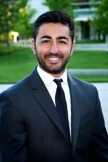
Alaa Bou Ghannam, MD
Dr. Bou Ghannam is a specialist in pediatric ophthalmology, adult strabismus, and neuro-ophthalmology. He is Clinical Instructor in the Department of Ophthalmology at the American University of Beirut Medical Center, Lebanon, and a Visiting Consultant at Moorfields Eye Hospital, Dubai, UAE.
Dr. Bou Ghannam completed his medical training and residency in Ophthalmology at the American University of Beirut, a fellowship in pediatric ophthalmology and adult strabismus at Children’s National Hospital and George Washington University, Washington, DC, and a fellowship in neuro-ophthalmology at the University of Colorado, Denver, CO, before he returned to Lebanon and the Middle East in 2017.
Dr. Bou Ghannam has a very active clinical practice and pursues clinical research in ophthalmology and neurology. He has published more than a dozen scientific papers and book chapters in leading medical journals. He is a member of the Lebanese Ophthalmological Society, North American Neuro-ophthalmology Society, and American Association for Pediatric Ophthalmology and Strabismus.
Abstract
Pediatric optic neuritis is a rare condition that can cause acute or subacute, temporary or permanent, vision loss in children. Compared to adults, optic neuritis in children is more likely to be bilateral and often follows a systemic infection or vaccination. It might also be a sequela of demyelinating diseases like Acute Disseminated Encephalomyelitis (ADEM), Multiple Sclerosis (MS) or Neuromyelitis Optica (NMO). Treatment is controversial but IV steroids is frequently used in the acute setting. Prognosis is usually excellent as most children recover full vision. This talk will discuss cases of pediatric optic neuritis and highlight the presentation, etiology, management and prognosis of this disease.
Optic Nerve Anomalies in Children

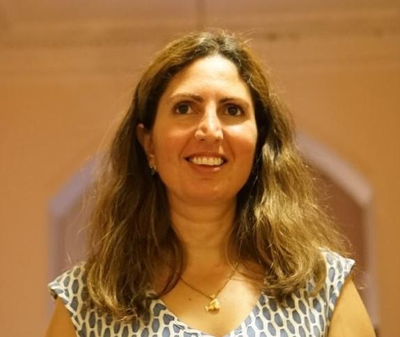
Mays El-Dairi, MD
Dr. Mays El-Dairi is an associate professor of pediatric ophthalmology and strabismus, and neuro-ophthalmology at Duke University. She completed her ophthalmology residency at the American University of Beirut, Lebanon, and fellowships in pediatric ophthalmology and strabismus, and neuro-ophthalmology at Duke University, Durham, NC. She joined the faculty at Duke in 2009.
Dr. El-Dairi is a clinician with expertise and interest in the areas of pediatric ophthalmology, neuro-ophthalmology and spectral domain optical coherence tomography (OCT) imaging. Her main research has been on the use of OCT imaging in children with a particular focus on evaluating the development of the pediatric optic nerve and retina in normal eyes, as well as in those with optic nerve cupping, papilledema, optic atrophy and variable neurologic injuries including hypoxic ischemic encephalopathy and complications of prematurity.
Dr. El-Dairi frequently organizes and participates in educational programs at the American Academy of Ophthalmology, the American Association for Pediatric Ophthalmology and Strabismus, the North American Neuro-ophthalmology Society and numerous Middle East region meetings.
Abstract
For children with anomalous optic nerve, three interventions need to be considered:
- A structural assessment to identify the type of abnormality (congenital vs. acquired), and the need for systemic workup (neuroimaging, endocrine, heart, kidney, hearing or other evaluations).
- A functional assessment to identify visual disability and need for accommodations.
- A follow-up plan depending on the risk of future vision loss or systemic involvement (risk for amblyopia, choroidal neovascularization, retinal detachment, glaucoma, hearing, cardiac or other systemic anomalies).
Sometimes, it is difficult to tell certain conditions apart, such as papilledema and pseudopapilledema, in which case more ophthalmic ancillary testing and closer follow up might be required. The role of the ophthalmologist is to work with the pediatric teams to decide upon the appropriate and cost-effective workup and follow-up.